Anatomy 3D Atlas

Brain anatomy in 3D:
isolated

Head Nervous system

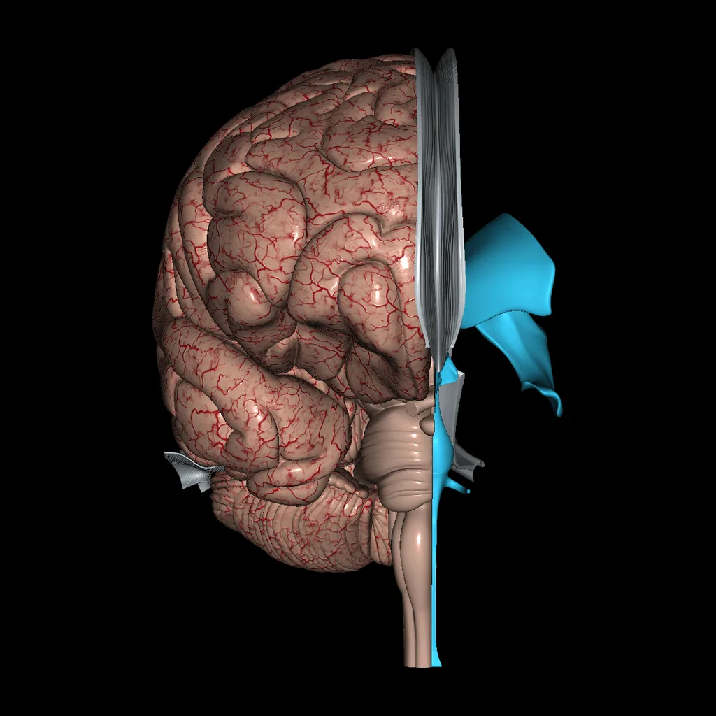
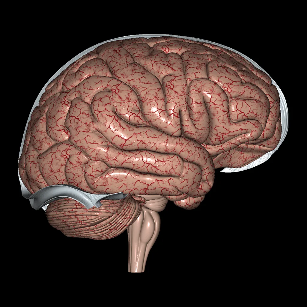
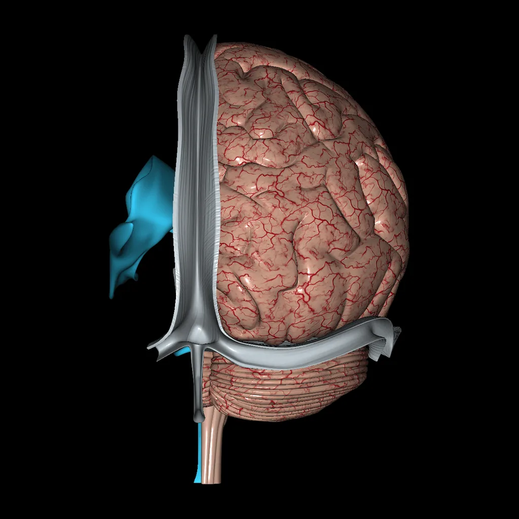
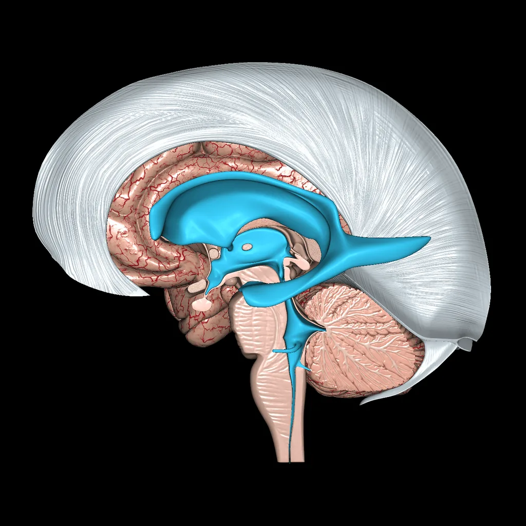
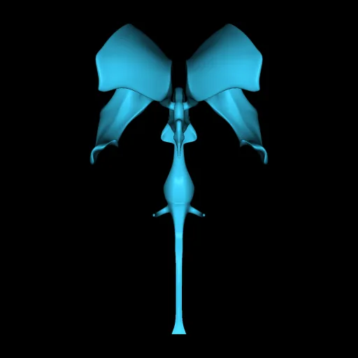
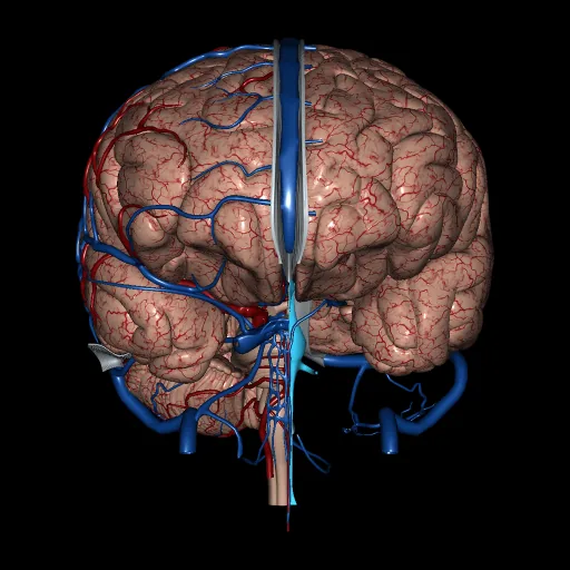
Brain (Isolated), anterior viewBrain (Isolated), lateral viewBrain (Isolated), posterior viewBrain (Isolated), medial view

The isolated brain, showing the cerebral hemispheres with their lobes, the cerebellum and the brainstem.

Each cerebral hemisphere is divided into frontal, parietal, temporal, occipital and insular lobes, separated by sulci such as the central and lateral sulci. The cerebellum coordinates movement and balance, and the brainstem, made up of the midbrain, pons and medulla oblongata, connects the brain to the spinal cord.

Systems in this model

  • Brain

Structures in this model (131)

Brain (131)
  • Right_Cerebral_hemisphere
  • Falx cerebri, Falx cerebelli, Tentorium cerebelli
  • Cerebellum (right)
  • Flocculus (right)
  • Ventricles
  • Brainstem (sectioned)
  • Corpus callosum
  • Fornix
  • Commisure of fornix
  • Caudate nucleus, Putamen
  • Amygdala
  • Globus pallidus lateral
  • Globus pallidus medial
  • Thalamus
  • Anterior commisure
  • Middle commisure (Interthalamic adhesion)
  • Choroid plexus (2° 3° ventricles)
  • Choroid plexus (4° ventricle)
  • Optic tract (CN II)
  • Frontal lobe
  • Parietal lobe
  • Temporal lobe
  • Occipital lobe
  • Limbic lobe
  • Superior parietal lobule
  • Inferior parietal lobule
  • Superior frontal gyrus
  • Middle frontal gyrus
  • Inferior frontal gyrus
  • Precentral gyrus
  • Postcentral gyrus
  • Superior temporal gyrus
  • Middle temporal gyrus
  • Inferior temporal gyrus
  • Supramarginal gyrus
  • Angular gyrus
  • Parahippocampal gyrus
  • Lateral occipitotemporal gyrus
  • Medial occipitotemporal gyrus
  • Uncus
  • Straight gyrus
  • Orbital gyrus
  • Cingulate gyrus
  • Cuneus
  • Medial frontal gyrus
  • Superior frontal sulcus
  • Inferior frontal sulcus
  • Lateral sulcus
  • Precentral sulcus
  • Central sulcus
  • Postcentral sulcus
  • Intraparietal sulcus
  • Parieto occipital sulcus
  • Calcarine sulcus
  • Tranverse occipital sulcus
  • Superior temporal sulcus
  • Inferior temporal sulcus
  • Preoccipital notch
  • Rhinal sulcus
  • Orbital sulcus
  • Olfactory sulcus
  • Cingulate sulcus
  • Marginal sulcus
  • Longitudinal cerebral fissure
  • Falx cerebri
  • Falx cerebelli
  • Tentorium cerebelli
  • Anterior lobe (Cerebellum)
  • Posterior lobe (Cerebellum)
  • Superior vermis
  • Inferior vermis
  • Nodule (Inferior vermis)
  • Tonsil (Cerebellum)
  • Primary fissure
  • Horizontal fissure
  • Pons
  • Middle cerebellar peduncle
  • Superior cerebellar peduncle
  • Cerebral peduncle (Cerebral crus)
  • Tuberculum cinereum
  • Posterior perforated substance (Interpenducular fossa)
  • Pineal body
  • Quadrigeminal plate (Superior colliculus)
  • Quadrigeminal plate (Inferior colliculus)
  • Superior medullary velum
  • Medulla oblongata
  • Pyramids (Brainsteam)
  • Olive
  • Cuneate tubercle
  • Gracile tubercle
  • Cuneate fasciculus
  • Gracile fasciculus
  • Septum pellucidum
  • Lamina terminalis
  • Hypophysis (pituitary gland)
  • Posterior commissure
  • Habenular commissure
  • Rostrum (Corpus callosum)
  • Medial geniculate body
  • Lateral geniculate body
  • Lateral ventricle (right)
  • Lateral ventricle (left)
  • Body of lateral ventricle
  • Anterior horn (Lateral ventricle)
  • Posterior horn (Lateral ventricle)
  • Inferior horn (Lateral ventricle)
  • Interventricular foramina (Monro)
  • 3rd ventricle
  • Suprapineal recess
  • Optic recess
  • Infundibulum
  • Cerebral aqueduct (Sylvius)
  • 4th ventricle
  • Lateral recess
  • Central canal
  • Genu (Corpus callosum)
  • Trunk (Corpus callosum)
  • Splenium (Corpus callosum)
  • Crus (Fornix)
  • Body (Fornix)
  • Column (Fornix)
  • Mammillary body
  • Hippocampus
  • Pes hippocampi
  • Taenia of fornix
  • Dentate gyrus
  • Head of Caudate nucleus
  • Body of Caudate nucleus
  • Tail of Caudate nucleus
  • Putamen
  • Pulvinar (Thalamic nuclei)