Anatomy 3D Atlas

Lower limb anatomy in 3D:
muscles and bones

Lower Limbs Musculoskeletal system

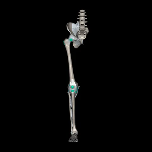
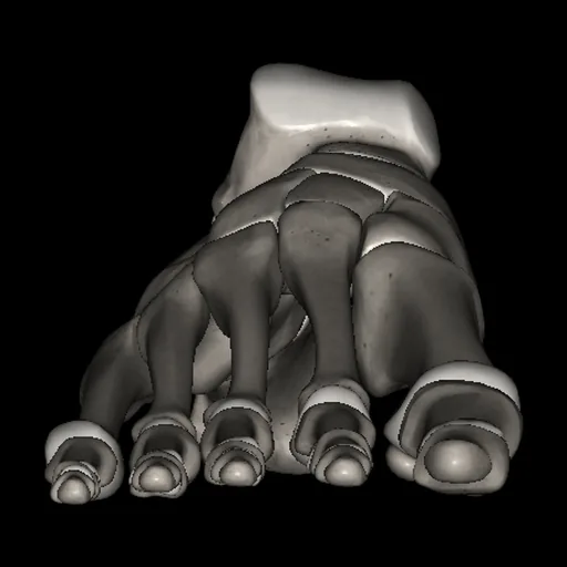
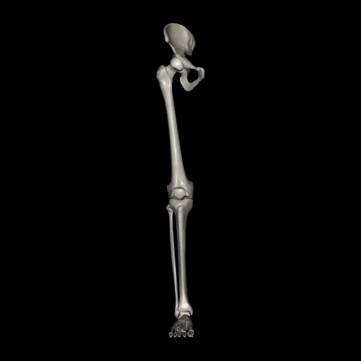
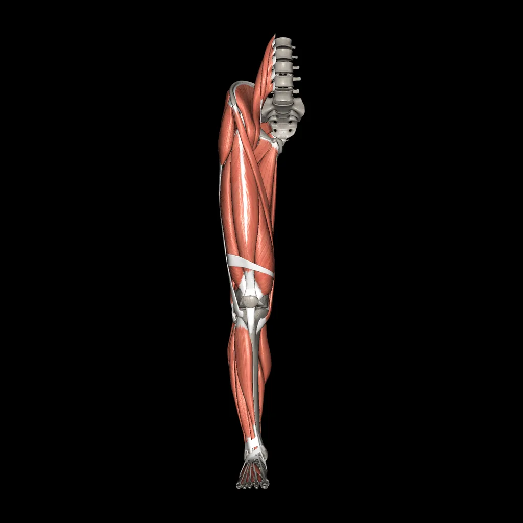
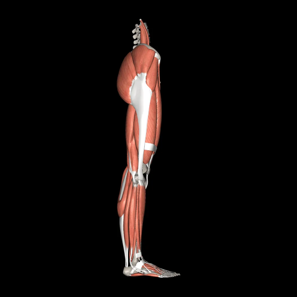
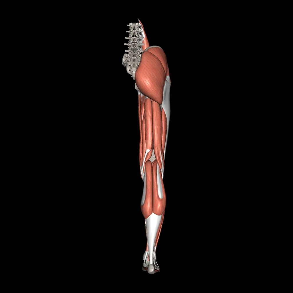
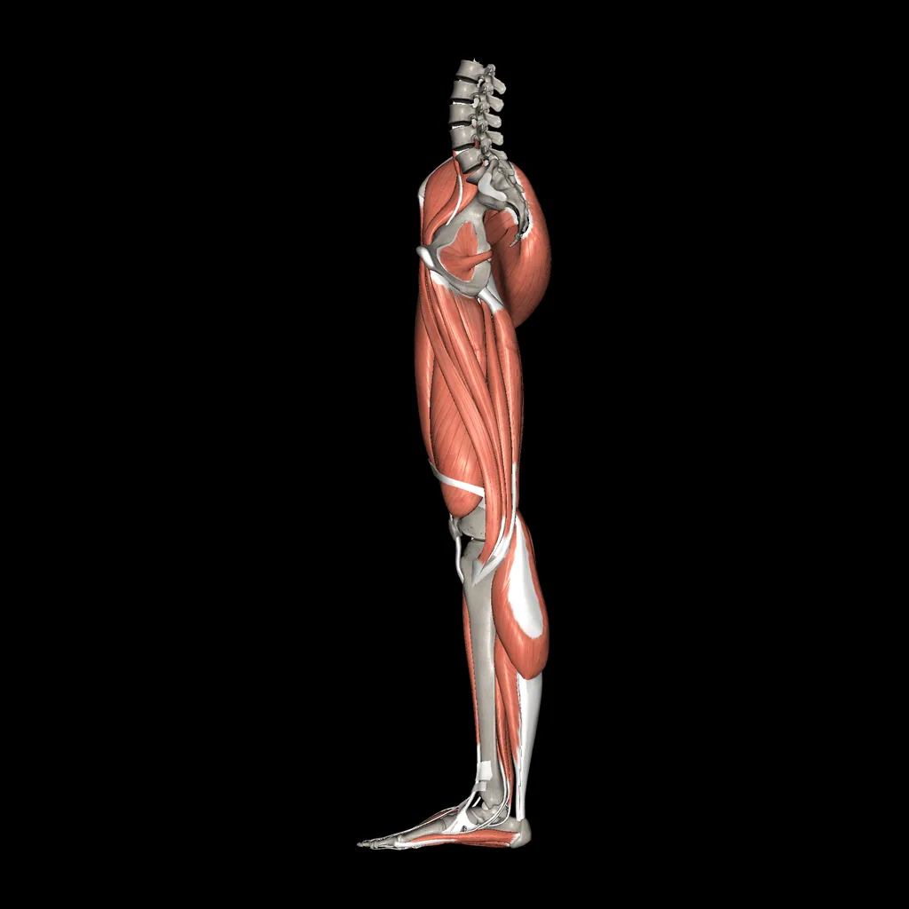
Lower limb (Muscles and bones), anterior viewLower limb (Muscles and bones), lateral viewLower limb (Muscles and bones), posterior viewLower limb (Muscles and bones), medial view

The bones of the lower limb with the muscles of the hip, thigh, leg and foot.

The gluteal muscles extend and abduct the hip, the anterior thigh compartment contains the quadriceps femoris, which extends the knee, and the posterior compartment the hamstrings. In the leg, the anterior compartment dorsiflexes the foot, the lateral compartment everts it and the posterior compartment, including gastrocnemius and soleus, plantarflexes it.

Systems in this model

  • Bones
  • Muscles

Structures in this model (279)

Bones (203)
  • Lumbar Vertebra, L1
  • Lumbar Vertebra, L2
  • Lumbar Vertebra, L3
  • Lumbar Vertebra, L4
  • Lumbar Vertebra, L5
  • Sacrum
  • Coccyx, 1
  • Coccyx, 2
  • Coccyx, 3
  • Coccyx, 4
  • Hip bone, Right
  • Femur, Right
  • Patella, Right
  • Tibia, Right
  • Fibula, Right
  • Talus, Right
  • Calcaneus, Right
  • Navicular, Right
  • Cuboid, Right
  • Lateral Cuneiform, Right
  • Intermediate Cuneiform, Right
  • Medial Cuneiform, Right
  • 1st Metatarsal, Right
  • 2nd Metatarsal, Right
  • 3rd Metatarsal, Right
  • 4th Metatarsal, Right
  • 5th Metatarsal, Right
  • Sesamoid, Right Foot
  • 1st Proximal Phalanx, Right Foot
  • 2nd Proximal Phalanx, Right Foot
  • 3rd Proximal Phalanx, Right Foot
  • 4th Proximal Phalanx, Right Foot
  • 5th Proximal Phalanx, Right Foot
  • 2nd Middle Phalanx, Right Foot
  • 3rd Middle Phalanx, Right Foot
  • 4th Middle Phalanx, Right Foot
  • 5th Middle Phalanx, Right Foot
  • 1st Distal Phalanx, Right Foot
  • 2nd Distal Phalanx, Right Foot
  • 3rd Distal Phalanx, Right Foot
  • 4th Distal Phalanx, Right Foot
  • 5th Distal Phalanx, Right Foot
  • Accessory process
  • Costal process
  • Inferior articular facet
  • Inferior articular process
  • Inferior vertebral notch
  • Intervertebral surface
  • Lamina of the vertebral arch
  • Mammillary process
  • Pedicle
  • Spinous process
  • Superior articular facet
  • Superior articular process
  • Superior vertebral notch
  • Vertebral body
  • Vertebral foramen
  • Anterior sacral foramen
  • Apex of sacrum
  • Auricular surface
  • Base of sacrum
  • Coccygeal cornu
  • Coccyx
  • Lateral sacral crest
  • Medial (Intermediate) sacral crest
  • Median sacral crest
  • Posterior sacral foramen
  • Promontory
  • Sacral canal
  • Sacral cornu
  • Sacral hiatus
  • Sacral tuberosity
  • Superior articular facet
  • Superior articular process
  • Transverse line
  • Tranverse process
  • Wing
  • Intervertebral foramen
  • Sacroiliac joint
  • Acetabular fossa
  • Acetabular margin
  • Acetabular notch
  • Acetabulum
  • Anterior gluteal line
  • Anterior inferior iliac spine
  • Anterior superior iliac spine
  • Arcuate line
  • Auricular surface
  • Body of ischium
  • Body of pubis
  • Gluteal surface
  • Greater sciatic notch
  • Iliac crest
  • Iliac fossa
  • Iliac tubercle
  • Iliac tuberosity
  • Iliac wing (Ala of ilium)
  • Iliopubic eminence
  • Inferior gluteal line
  • Inferior pubic ramus
  • Inner lip
  • Ischial spine
  • Ischial tuberosity
  • Lesser sciatic notch
  • Lunate surface
  • Obturator foramen
  • Outer lip
  • Pectineal line (Pecten pubis)
  • Posterior inferior iliac spine
  • Posterior superior iliac spine
  • Pubic tubercle
  • Ramus of ischium
  • Superior pubic ramus
  • Symphyseal surface
  • Adductor tubercle
  • Fovea of femur head
  • Gluteal tuberosity
  • Greater trochanter
  • Head of femur
  • Intercondylar fossa
  • Intercondylar line
  • Intertrochanteric crest
  • Intertrochanteric line
  • Lateral condyle
  • Lateral epicondyle (femur)
  • Lateral supracondylar line
  • Lesser trochanter
  • Linea aspera (lateral lip)
  • Linea aspera (medial lip)
  • Medial condyle
  • Medial epicondyle (femur)
  • Medial supracondylar line
  • Neck of femur
  • Patellar surface
  • Pectineal line
  • Popliteal surface
  • Shaft of femur
  • Trochanteric fossa
  • Anterior surface
  • Apex of patella
  • Base of patella
  • Lateral articular facet
  • Medial articular facet
  • Anterior border
  • Articular facet of medial melleolus
  • Fibular articular facet
  • Fibular notch
  • Inferior articular surface
  • Intercondylar eminence
  • Interosseous border
  • Lateral condyle of tibia
  • Lateral surface
  • Malleolar sulcus
  • Medial border
  • Medial condyle of tibia
  • Medial malleolus
  • Medial surface
  • Nutrient foramen
  • Oblique line
  • Posterior surface
  • Soleal (popliteal) line
  • Superior articular surface (medial facet)
  • Superior articular surface (lateral facet)
  • Tibial tuberosity
  • Anterior border
  • Apex
  • Articular facet for tibia
  • Head of Fibula
  • Interosseous border
  • Articular surface of lateral malleolus
  • Lateral malleolus
  • Lateral surface
  • Malleolar fossa
  • Medial crest
  • Medial surface
  • Neck of fibula
  • Posterior border
  • Posterior surface
  • Talocrural (Mortise) joint
  • Base of 1st metatarsal
  • Base of 2nd proximal phalanx
  • Body of 1st metatarsal
  • Body of 2nd proximal phalanx
  • Calcaneal tuberosity
  • Groove for fibularis (peroneus) longus tendon
  • Head of 1st metatarsal
  • Head of 2nd proximal phalanx
  • Head of talus
  • Lateral process of calcaneal tuberosity
  • Lateral tubercle of talus
  • Medial process of calcaneal tuberosity
  • Medial tubercle of talus
  • Neck of talus
  • Peroneal (Fibular) trochlea
  • Subtalar joint
  • Sustentaculum tali
  • Tarsal sinus
  • Tarsometatarsal joint
  • Transverse tarsal joint
  • Trochlea of talus
  • Tuberosity of 5th metatarsal
  • Tuberosity of cuboid
  • Tuberosity of navicular
Muscles (76)
  • Iliacus (Iliopsoas)
  • Psoas major (Iliopsoas)
  • Psoas minor
  • Gluteus maximus
  • Gluteus medius
  • Gluteus minimus
  • Tensor fasciae latae
  • Iliotibial tract
  • Piriformis
  • Obturator internus
  • Obturator externus
  • Gemellus superior
  • Gemellus inferior
  • Quadratus femoris
  • Sartorius
  • Quadriceps femoris, Rectus
  • Quadriceps femoris, Vastus lateralis
  • Quadriceps femoris, Vastus intermedius
  • Quadriceps femoris, Vastus medialis
  • Pectineus
  • Adductor longus
  • Adductor brevis
  • Adductor magnus
  • Adductor minimus
  • Gracilis
  • Biceps femoris
  • Semitendinosus
  • Semimembranosus
  • Articularis Genus
  • Arciform fascia
  • Patellar ligament
  • Tibialis anterior
  • Extensor digitorum longus
  • Fibularis longus (Peroneus longus)
  • Fibularis brevis (Peroneus brevis)
  • Fibularis tertius (Peroneus tertius)
  • Extensor hallucis longus
  • Triceps surae, Gastrocnemius, Lateral and Medial head
  • Triceps surae, Soleus
  • Plantaris
  • Popliteus
  • Tibialis posterior
  • Flexor digitorum longus
  • Flexor hallucis longus
  • Extensor hallucis brevis
  • Extensor digitorum brevis
  • Abductor hallucis
  • Flexor hallucis brevis
  • Adductor hallucis, Transverse head
  • Adductor hallucis, Transverse head
  • Adductor hallucis, Transverse head
  • Adductor hallucis Oblique head
  • Abductor Digiti Minimi
  • Opponens Digiti minimi
  • Flexor Digiti Minimi Brevis
  • Flexor digitorum brevis
  • Quadratus plantae; Flexor accessorius
  • Lumbrical 1st (foot)
  • Lumbrical 2nd (foot)
  • Lumbrical 3rd (foot)
  • Lumbrical 4th (foot)
  • Dorsal interosseous 1st (bipennate) (foot)
  • Dorsal interosseous 2nd (bipennate) (foot)
  • Dorsal interosseous 3rd (bipennate) (foot)
  • Dorsal interosseous 4th (bipennate) (foot)
  • Plantar interosseous 1st
  • Plantar interosseous 2nd
  • Plantar interosseous 3rd
  • Superior extensor retinaculum
  • Inferior extensor retinaculum
  • Long plantar ligament
  • Biceps femoris, long head
  • Biceps femoris, short head
  • Gastrocnemius, lateral head
  • Gastrocnemius, medial head
  • Calcaneal tendon (Achilles tendon)