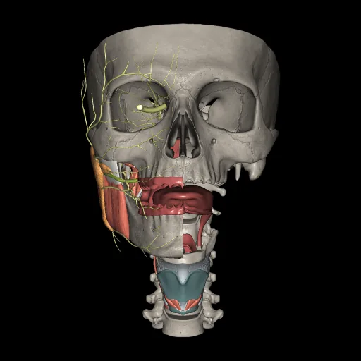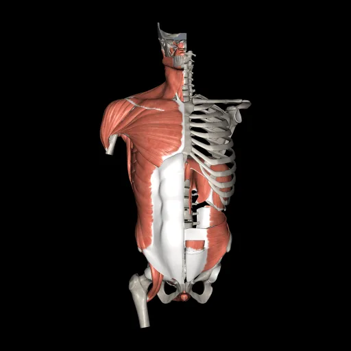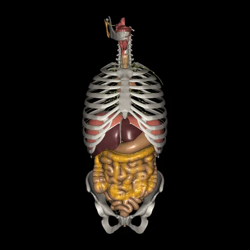
Digestive system anatomy in 3D:
isolated
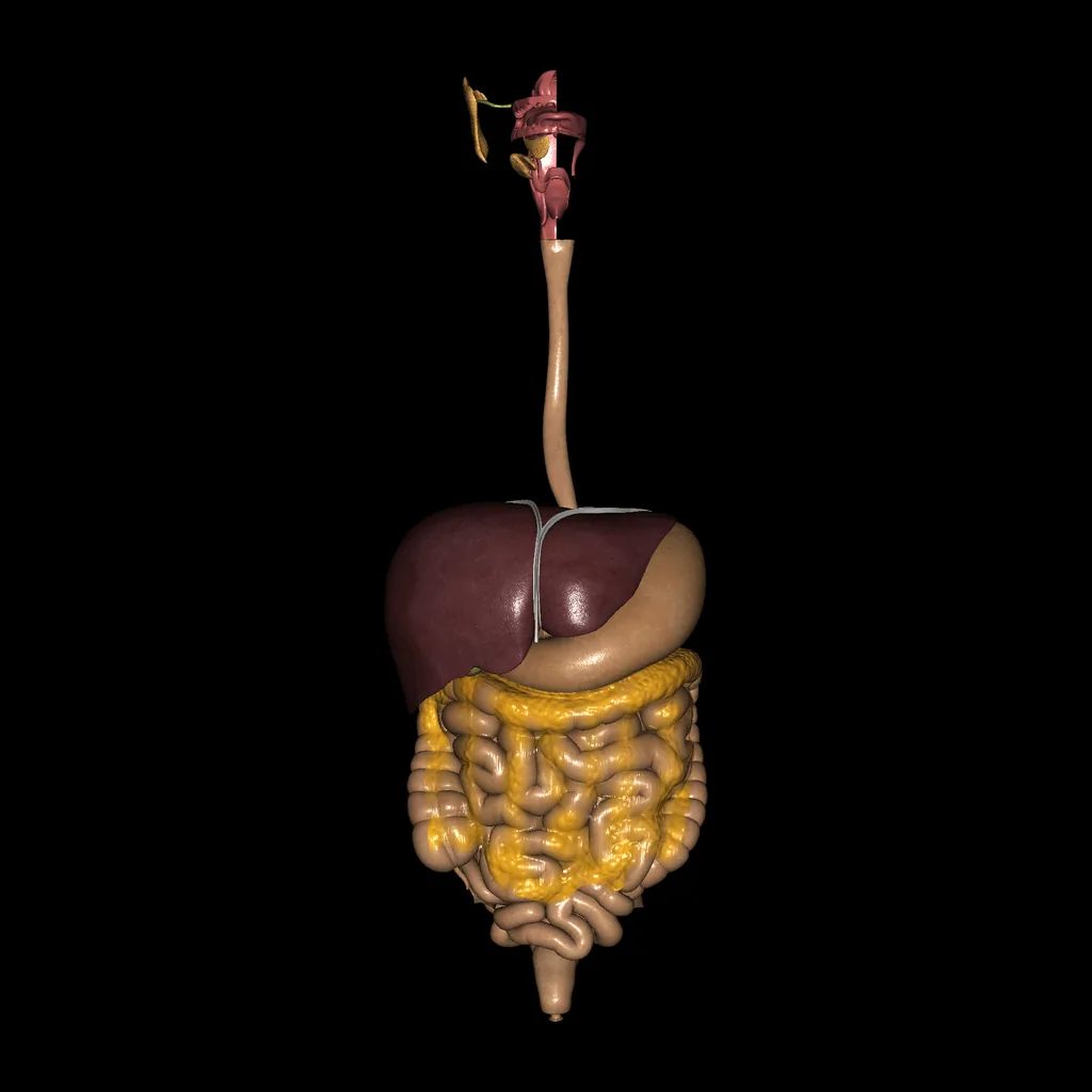
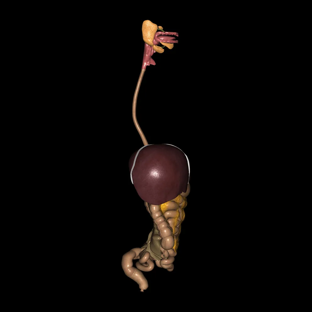
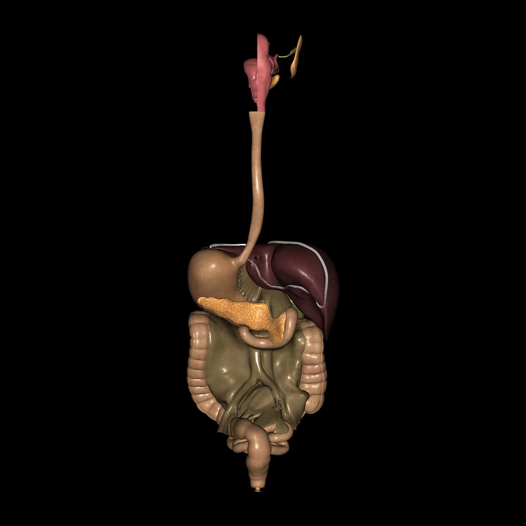
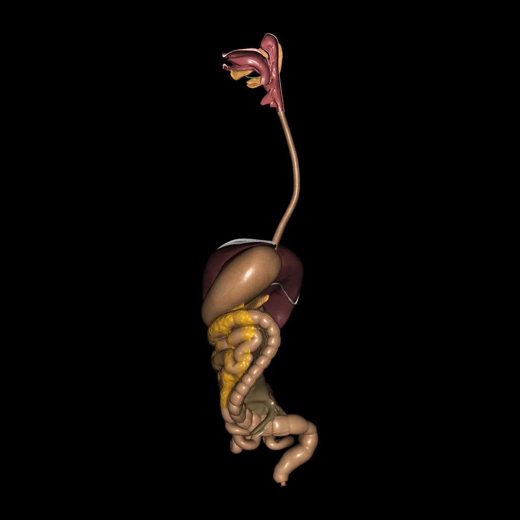
The isolated digestive system: oesophagus, stomach, intestines, liver, gallbladder and pancreas.
The stomach is divided into cardia, fundus, body and pyloric part, and the small intestine into duodenum, jejunum and ileum. The large intestine includes the caecum with the appendix, the ascending, transverse, descending and sigmoid colon, the rectum and the anal canal.
Systems in this model
- Digestive
Structures in this model (142)
Digestive system (142)
- Nasopharynx, Oropharynx, Laryngopharinx
- Parotid Gland
- Accessory Parotid Gland
- Parotid duct
- Sublingual gland
- Submandibular gland
- Submandibular duct (Wharton duct)
- Tongue
- Esophagus
- Stomach
- Duodenum
- Pancreatic duct
- Accessory pancreatic duct
- Pancreas
- Liver
- Liver ligaments
- Gallbladder
- Lesser omentum
- Greater omentum
- Mesentery,Transverse Mesocolon and Sigmoid Mesocolon
- Small intestine
- Large intestine (Colon)
- Nasopharynx
- Oropharynx
- Laryngopharinx
- Choanae
- Pharyngeal tonsil
- Torus tubarius
- Pharyngeal opening of pharygotympanic tube
- Pharyngeal recess
- Torus levatorius
- Hard palate
- Soft palate
- Uvula
- Palatine tonsil
- Palatine glands
- Inlet of larynx (aditus)
- Palatoglossal arch
- Palatopharyngeal arch
- Interarytenoid notch
- Piriform recess
- Dorsum of tongue
- Apex of tongue
- Body of tongue
- Root of tongue
- Filiform paillae
- Vallate papillae
- Foliate papillae
- Fungiform papillae
- Median sulcus
- Lingual tonsil
- Sublingual caruncle
- Frenulum of tongue
- Foramen cecum
- Sulcus terminalis
- Esophagus
- Adventitia
- Muscle coat: longitudinal fibers
- Muscle coat: circular fibers
- Mucosa: longitudinal folds
- Esophageal hiatus
- Cardia
- Fundus
- Body
- Pyloric antrum
- Pyloric canal
- Pyloric orifice
- Greater curvature (Stomach)
- Lesser curvature (Stomach)
- Cardial notch
- Angular notch
- Head of pancreas
- Body of pancreas
- Tail of pancreas
- Pancreatic notch
- Right lobe of liver
- Left lobe of liver
- Caudate lobe
- Quadrate lobe
- Fossa for gallbladder
- Colic impression
- Renal impression
- Gastric impression
- Esophageal impression
- Suprarenal impression
- Duodenal impression
- Bare area
- Falciform ligament
- Coronary ligament
- Coronary ligament (hepatorenal)
- Right triangular ligament
- Left triangular ligament
- Round ligament
- Hepatoduodenal ligament
- Hepatogastric ligament
- Ascending mesocolon
- Transverse mesocolon
- Descending mesocolon
- Mesoappendix
- Gallbladder
- Common bile duct
- Common hepatic duct
- Infundibulum
- Right hepatic duct
- Left hepatic duct
- Cystic duct
- Fundus (Gallbludder)
- Neck (Gallbludder)
- Body (Gallbludder)
- Duodenum
- Jejunum
- Duodenoljejunal flexure
- Superior duodenal flexure
- Terminal part of ileum
- Frenulum
- Ileocecal orifice
- Ileocecal lips
- Ileum
- Cecum
- Vermiform appendix
- Haustra
- Descending colon
- Transverse colon
- Ascending colon
- Sigmoid colon
- Right colic flexure
- Left colic flexure
- Free taenia
- Omental taenia
- Mesocolic taenia
- Semilunar folds
- Rectosigmoid junction
- Superior transverse fold
- Middle transverse fold
- Inferior transverse fold
- Rectal ampulla
- Anorectal line
- Anal columns (Margagni)
- Anal sinuses
- Anal valves
- Pecten
- Anoderm
Related Models
A list of the most relevant models related to Digestive system (Isolated)